By the time you see it,
it is already late
Retinal disease gives almost no early warning, so when a patient notices it the disease has often progressed.
A retina specialist trained at Severance and Oxford finds it before you feel it.
The retina is the film
inside your eye
The retina is a thin layer of neural tissue that receives light, converts it into signals and sends them to the brain through the optic nerve. At its centre sits the macula, which does most of the work when you look at something directly.
Once damaged, retinal tissue rarely recovers. That is why finding it early and treating it precisely matters more here than almost anywhere else in the eye.
Conditions we treat
The earlier these are found, the more vision can be kept.
Macular degeneration
The centre of what you look at becomes blurred or distorted, and reading becomes difficult.
Diabetic retinopathy
Long-standing diabetes damages retinal vessels, which leak or close, and vision falls without warning.
Retinal detachment
Part of the visual field is suddenly covered as if by a curtain. This is an emergency.
Floaters
Spots, threads or cobwebs drifting across vision. Often benign, sometimes a warning sign.
Retinal vascular disease
A retinal vessel blocks or bleeds, causing a sudden loss of vision.
Vitreous disease
The vitreous clouds or pulls on the retina, producing a range of symptoms.
A sudden increase in floaters, flashes of light, or a shadow over part of your vision may indicate retinal detachment. Do not wait for an appointment. Call +82-2-553-3588.
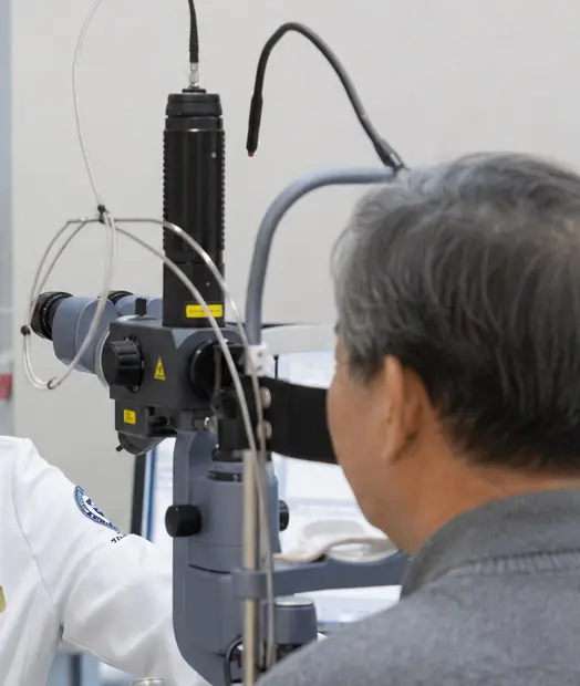
Not every ophthalmologist
treats the retina
Retina and vitreous work requires more than board certification in ophthalmology.
It requires a separate fellowship before a doctor can manage and operate on these conditions.
Every retina specialist is an ophthalmologist. Not every ophthalmologist is a retina specialist.
About the doctorTreatment given
before symptoms start
Were you told at a health check that diabetic retinopathy is suspected?
Has laser been suggested while your eyes still feel completely normal?
Retinal laser is not a treatment for pain. It is preventive work done while there are no symptoms, sealing abnormal vessels and damaged areas to stop the disease from advancing.
- Performed in the outpatient clinic, usually ten to twenty minutes
- Normal daily activity is possible the same day
- Reduces the risk of complications and slows progression
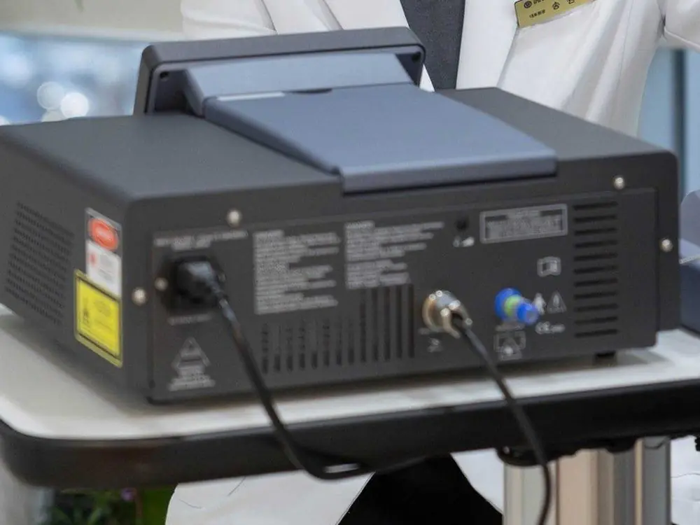
Imaging and diagnosis
Equipment of the standard used in university hospitals, with imaging and reading completed in a single visit.
Optical coherence tomography
A high resolution cross section of the retina, showing oedema and change that the surface does not reveal.
Fundus photography
A wide area of the retina captured at once, including the periphery.
Fluorescein angiography
Dye imaging of the retinal vessels to locate leakage.
Ocular measurement
Precise calculation of intraocular lens power before cataract surgery.
Visual field testing
Field loss recorded numerically so progression can be judged over time.
Slit lamp microscopy
Magnified examination of the cornea, lens and ocular surface.
Which tests are performed depends on your symptoms and diagnosis. Not every test is done at every visit.
Frequently asked
How long does the examination take?
Do floaters always need treatment?
I have diabetes but my eyes feel fine. Do I need a retinal exam?
Can macular degeneration be cured?
Is retinal laser treatment painful, and how long does it take?
Damaged retina
rarely comes back
If you have symptoms, do not put off a proper examination.
Imaging, reading and diagnosis are finished on the day you visit.
A retinal examination requires dilating drops. You should not drive yourself for three to four hours afterwards.